A Guide to Common Orthopaedic Procedures
TPLO (Tibial Plateau Levelling Osteotomy)
Understanding Cranial Cruciate Ligament Injuries
In dogs, the cranial cruciate ligament (CCL) plays a crucial role in stabilizing the knee (stifle) joint. When this ligament is damaged or ruptured, it leads to instability, causing pain and lameness. This condition is akin to anterior cruciate ligament (ACL) injuries in humans.
What is TPLO Surgery?
Tibial Plateau Levelling Osteotomy (TPLO) is a surgical procedure designed to address CCL injuries by altering the biomechanics of the knee joint. Instead of replacing the damaged ligament, TPLO changes the angle of the tibial plateau (the top part of the shinbone) to stabilize the joint during weight-bearing activities.
The Surgical Procedure
Preoperative Assessment: Detailed radiographs are taken to measure the existing angle of the tibial plateau and plan the surgical approach.
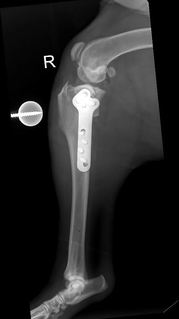
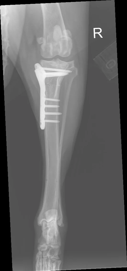
Surgical Technique: Under general anaesthesia, a curved cut (osteotomy) is made in the top portion of the tibia. This segment is then rotated to achieve a more level orientation, typically aiming for an angle between 5 to 6 degrees.
Stabilization: The repositioned bone is secured using a specially designed metal plate and screws, ensuring stability as the bone heals in its new alignment.
Benefits of TPLO
Enhanced Stability: By modifying the tibial plateau angle, TPLO eliminates the need for the CCL, providing inherent stability to the knee joint.
Rapid Recovery: Many dogs begin bearing weight on the operated leg within days post-surgery, facilitating a quicker return to normal activities.
Long-Term Success: TPLO has a high success rate, especially in large or active breeds, and reduces the progression of osteoarthritis compared to other surgical methods.
Postoperative Care and Recovery
Recovery from TPLO surgery typically spans 8 to 12 weeks. During this period, controlled exercise and adherence to postoperative guidelines are essential. Follow-up radiographs are usually performed around 6 weeks post-surgery to assess bone healing. Most dogs regain full function and return to their normal lifestyle and exercise regime once their recovery is complete.
Total Hip Replacement (THR)
Understanding Canine Hip Dysplasia and Osteoarthritis
Canine hip dysplasia is a developmental condition where the hip joint doesn’t form properly, leading to looseness and instability. Over time, this can result in chronic pain, inflammation, and secondary osteoarthritis (OA). Dogs with OA may show signs such as stiffness, difficulty rising, reduced activity, and reluctance to jump or climb stairs.
In more advanced cases, especially when medical management no longer provides adequate relief, Total Hip Replacement (THR) may be the most effective option to restore comfort and mobility.
What is Total Hip Replacement?
Total Hip Replacement is a surgical procedure that replaces the damaged hip joint with a prosthetic implant. It involves removing both the ball (femoral head) and socket (acetabulum) components of the joint and replacing them with artificial parts, restoring smooth, pain-free movement.
THR is considered the gold standard for end-stage hip conditions, including:
Severe hip dysplasia
Chronic subluxation or dislocation
Advanced osteoarthritis of the hip joint
The Surgical Procedure:
Preoperative Assessment: A full workup including physical examination, bloodwork, and radiographs ensures the dog is a suitable candidate for THR.
Anaesthesia and Preparation: Under general anaesthesia, the hip area is surgically prepared.
Joint Access and Removal: The surgeon carefully removes the diseased femoral head and reshapes the acetabulum.
Implant Placement: Specialized prosthetic components are securely fitted into the bone (using either cemented or cementless techniques) depending on the case.
Closure and Recovery: Soft tissues are sutured, and the dog is closely monitored during recovery.
Benefits of THR:
Eliminates Pain: Removes the source of chronic hip discomfort.
Restores Normal Function: Allows dogs to walk, run, and play without restriction.
Ideal for Severe Osteoarthritis: Particularly beneficial for dogs with advanced degenerative joint disease who are no longer responsive to conservative treatments.
High Success Rate: Over 90% of dogs regain excellent limb function and quality of life.
Recovery and Postoperative Care:
Hospital Stay: Typically 1–3 days.
Rest and Restriction: Limited activity for 6–8 weeks with short, controlled leash walks.
Rehabilitation: Gradual reintroduction of movement and muscle strengthening.
Follow-Up: Post-op radiographs and examinations ensure proper healing and implant stability.
Potential Risks and Considerations:
While THR is highly successful, potential risks include:
Implant loosening or dislocation
Surgical site infection
Rare nerve injury
Close postoperative monitoring and adherence to recovery protocols greatly reduce these risks.

Patellar Luxation
Understanding Patellar Luxation
Patellar luxation is a condition where the kneecap (patella) dislocates from its normal position within the groove of the thigh bone (femur). This displacement can lead to pain, lameness, and reduced mobility in affected animals.
Causes and Risk Factors
Patellar luxation can result from congenital anatomical defects, trauma, or a combination of both. Certain breeds, particularly small and toy breeds, are more predisposed to this condition. The condition is graded based on severity, ranging from Grade I (mild) to Grade IV (severe), which helps determine the appropriate treatment approach.
Symptoms
Dogs with patellar luxation may exhibit:
Intermittent lameness or skipping gait
Sudden lifting of a hind limb while walking
Reluctance to jump or run
Signs of discomfort or pain in the knee area
In some cases, the condition may be present in both hind limbs, leading to a more noticeable impact on mobility.
Diagnosis
A thorough physical examination by a veterinarian is essential. In some cases, imaging techniques like X-rays may be employed to assess the severity and plan appropriate treatment.
Treatment Options
Non-Surgical Management:
Weight management to reduce stress on joints
Physical therapy to strengthen surrounding muscles
Pain relief medications and joint supplements
Surgical Intervention:
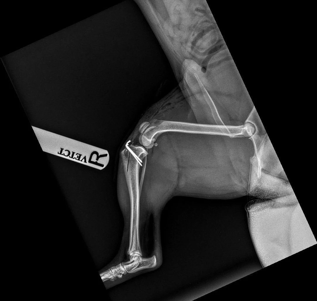
In moderate to severe cases, surgical correction may be recommended. Procedures can include realignment of the patella, deepening of the femoral groove, or soft tissue adjustments to stabilize the kneecap. The specific surgical approach depends on the individual dog's condition and the severity of the luxation. Patellar luxation is commonly regarded as a condition that can be corrected with relative ease; however, achieving the best possible outcome requires careful planning and precise execution. Selecting the appropriate surgical technique is crucial, as the wrong approach—or poor execution—can lead to further complications. Treatment often involves a combination of procedures, such as deepening the femoral trochlear groove where the kneecap sits and adjusting the alignment of the patellar tendon by repositioning the tibial crest. The latter is especially critical for preventing recurrence, yet it can sometimes be overlooked. In more complex cases, realigning the bones through corrective osteotomy of the femur or tibia may be necessary, such as in case of DFO or distal femoral osteotomy. In rare instances, where the femoral trochlea is significantly damaged, a partial joint replacement may be the only viable option (PGR or patella groove replacment). Making these decisions often depends on the surgeon’s experience and may require advanced imaging, such as a CT scan.
Prognosis
With appropriate treatment, many dogs recover well and return to normal activities. Early intervention can prevent the progression of joint damage and improve the quality of life.
Angular limb deformity
Understanding angular limb deformities (ALD)
Sometimes, the bone growth with an abnormal twist. The subsequent abnormal weight bearing over the joints leads progressivevely to pain (limp) and arthritis. Sometimes this can also happen when a broken bone heals at an abnormal angle.
Causes and Risk Factors
Small breed such a Shitzu and generally all short legged breeds are predisposed, often to a deformity of the front limbs known as “radius curvus”. Mor particualrly Dachshund are predisposed to a specific abnormal growth of their back legs known as “pes varus”. Large breeds are more predisposed to growth issues following a break or a trauma, particularly if they are very young.
Symptoms
Dogs with ALD
Intermittent , progressively worsening lameness
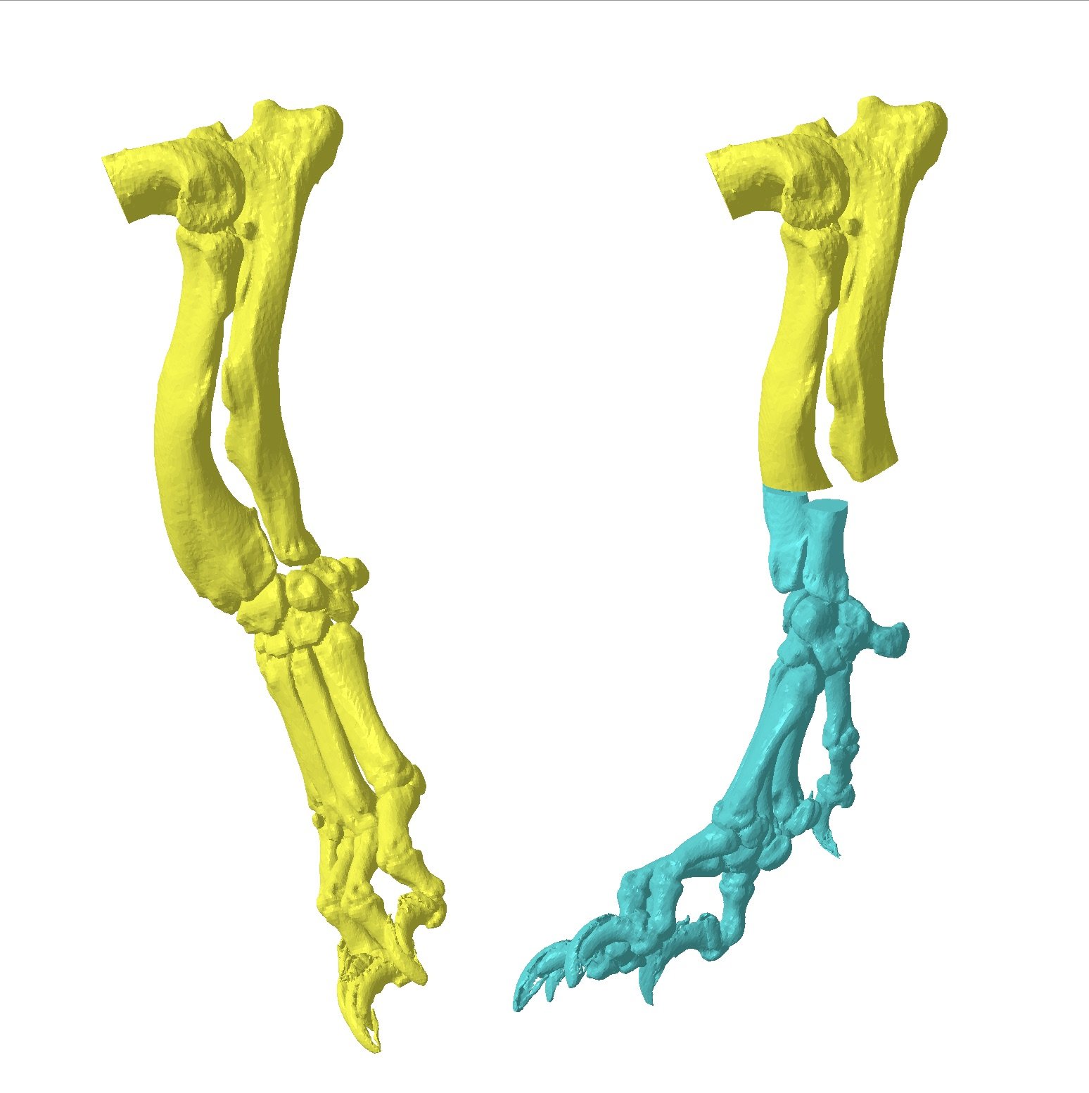
limb sticking markedly outwards
visibel assymetry between the left and right limb
Abnormal leg movement during the walk.
Diagnosis
A thorough physical examination by a veterinarian is essential. In some cases, imaging techniques like X-rays or preferable CT scan may be employed to assess the severity and plan appropriate treatment.
Treatment Options
Non-Surgical Management:
This is only possible when the deformity is not creating any lameness. In such case, the angular deformity is just esthetical and treating it is not possible. In case of lameness, you may consider pain relief and possibly a brace, however, severe cases will not respond very well. It is also important to understand that the longer the condition is left untreated surgically (from the moment you rnotice the lameness), the more likely the progression of the arthritis and with this, a lesser chance to have a good long term outcome.
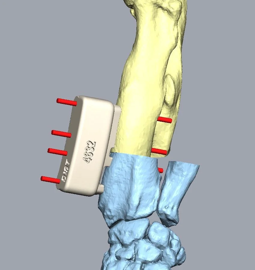
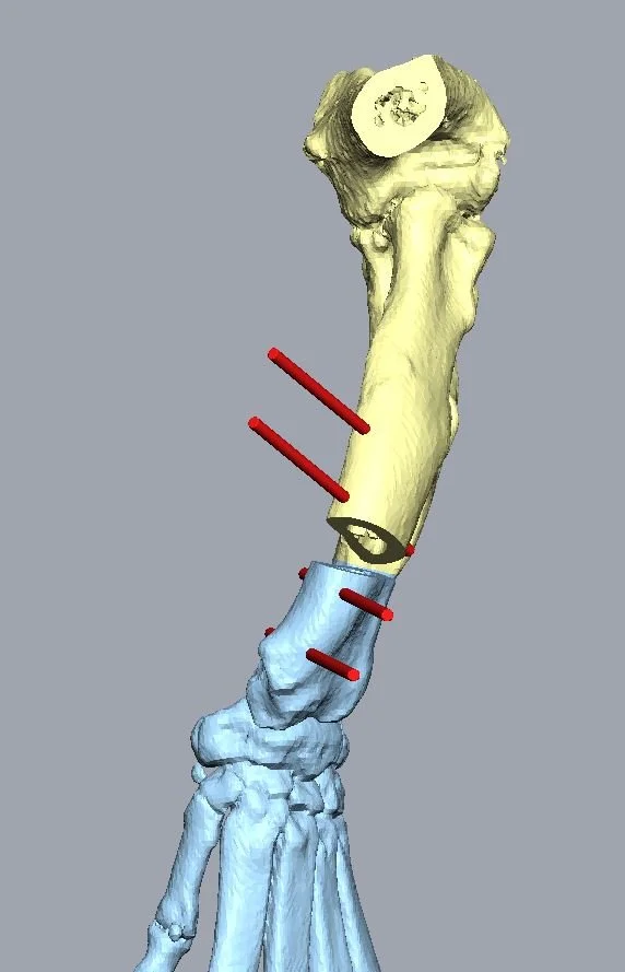
Surgical Intervention:
There are various ways to deal with these conditions, the most successful cases are those treatted early on, and for which the deformity is assessed fully and corrected appropriately. The most recent techniques of 3D printing, bone modelling and design of customised surgical guides allow an increadible precision and planning for those procedures. Here at Gainsborough referrals, we can offer those state of art procedures from £5000.
Prognosis
With appropriate treatment, many dogs recover well and return to normal activities. Early intervention can prevent the progression of joint damage and improve the quality of life.